The magnetic resonance (magneTIc resonance, MR) phenomenon was discovered independently in 1945 by the Stanford team led by Bullock and the MIT team led by Purcell. But it was not until the birth of spectrometers with high magnetic fields, high resolutions, and Fourier transform technology in the 1960s that the application of magnetic resonance in the field of biology made substantial progress.
In recent years, due to the advantages of high contrast, high resolution, no dead angle observation, and no side effects on the human body, magnetic resonance imaging has attracted a large number of researchers to invest in research, which has made great progress in the following aspects:
Echo plane imaging (echoplannar maging, EPI) greatly shortens the imaging time of MR, and high-resolution images can be obtained within 100-200ms (pixel width <1.5mm =. Lower resolution images (pixel width> 3mm ) It takes only 50ms.
Magnetic resonance angiography (magneTIc esonance angiography, MRA) does not require contrast agents to obtain angiographic images, which is superior to CT and X-ray angiography. There are also magnetic resonance perfusion and penetration weighted imaging, which not only provide information on the morphology of human tissues and organs, but also provide information on functions.
Magnetic resonance imaging intervention, with good tissue contrast, can accurately distinguish the interface of the lesion and determine the target; sub-millimeter spatial resolution facilitates lesion positioning and intervention guidance; multi-layer and three-dimensional spatial imaging allows comprehensive observation of important anatomical structures ; Fast and ultra-fast imaging sequences enable near real-time observation of changes in physiological movements, interventional instruments, and interventions.
Techniques for eliminating artifacts, such as spatial pre-saturation, gradient magnetic moment balancing and fast imaging, can effectively eliminate magnetic resonance images caused by human body physiological movements such as breathing, blood flow, cerebrospinal fluid pulsation, heart beat, gastrointestinal peristalsis, etc. Artifacts.
The following figure is a block diagram of a general magnetic resonance system:
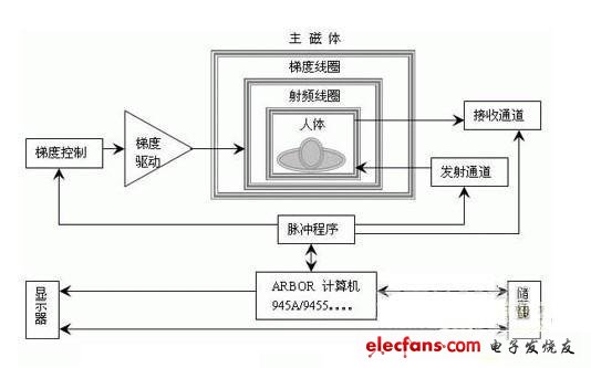
Figure General magnetic resonance system block diagram
The main magnet of the magnetic resonance imaging system is used to generate a highly uniform and stable static magnetic field, which can be permanent, normal and superconducting magnets. Generally, the main magnet is made into a cylindrical or rectangular cavity, in which not only the coil of the main magnet, but also the coil of the gradient magnetic field in the X, Y, and Z directions, as well as the RF transmission coil and receiving coil of the whole body can be installed. among them.
The gradient generator generates a gradient current with a certain switch shape, which is amplified and sent to the gradient coil by the driving circuit to generate the required gradient magnetic field.
The radio frequency transmitter includes a frequency synthesizer, RF formation, amplification and power amplifier, and generates the required radio frequency pulse current and sends it to the radio frequency transmission coil.
The receiver consists of instruments such as front-end, radio frequency, band-pass filtering, detection, low frequency and A / D conversion. The received magnetic resonance signal is amplified and processed into a digital signal and enters the computer.
The computer reconstructs the collected data, and sends the image data to the display for display. In addition, the computer is also responsible for controlling the operation of each part of the entire system, so that the actions of each part of the entire imaging process are coordinated and consistent, and produce the required high-quality images.
Since the computer is the control center of the system, its computing power and stability are particularly important. The ARBOR computer is widely used in many medical devices due to its powerful functions, stable performance, and strong environmental adaptability. EmETXe-i9455 has been successfully applied to large-scale superconducting magnetic resonance imaging systems.
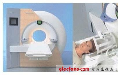
In order to accelerate the research and popularization of magnetic resonance imaging technology, many developers have developed and produced many portable ultra-small magnetic resonance imagers. ARBOR EmETXe-i9455 has been successfully used in ultra-small magnetic resonance imaging instruments with its small size, strong computing power, low power consumption, and stable performance. This type of equipment is mainly used for local lesion examination in hospitals and college teaching or spectroscopy research.
Square Hole Breadboard,400 Tie Point Breadboard,Makeronics Breadboard,Small Solderless Breadboard
Cixi Zhongyi Electronics Factory , https://www.zypcbboards.com